Archivo:Variant Creutzfeldt-Jakob disease (vCJD), H&E.jpg
No se dispone de una resolución más alta.
Variant_Creutzfeldt-Jakob_disease_(vCJD),_H&E.jpg (700 × 554 píxeles; tamaño de archivo: 80 kB; tipo MIME: image/jpeg)
,_H%26E.jpg?uselang=es)
Resumen
| Descripción |
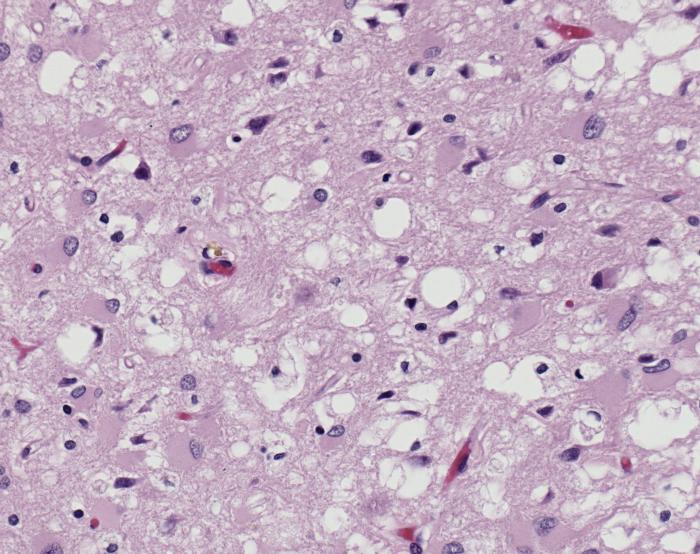
ID#: 10131 Magnified 100X, and stained with H&E (hematoxylin and eosin) staining technique, this light photomicrograph of brain tissue reveals the presence of prominent spongiotic changes in the cortex, and loss of neurons in a case of variant Creutzfeldt-Jakob disease (vCJD). Variant Creutzfeldt-Jakob disease (vCJD) is a prion disease that was first described in 1996 in the United Kingdom. There is now strong scientific evidence that the agent responsible for the outbreak of prion disease in cows, bovine spongiform encephalopathy (BSE or 'mad cow' disease), is the same agent responsible for the outbreak of vCJD in humans. Both disorders are invariably fatal brain diseases with unusually long incubation periods measured in years, and are caused by an unconventional transmissible agent called a prion. vCJD is not the same disease as classic CJD. It has different clinical and pathologic characteristics from classic CJD. Each disease also has a particular genetic profile of the prion protein gene. |
| Fuente | Public Health Image Library (PHIL) ID#: 10131 |
| Autor |
Content Providers(s): CDC/ Teresa Hammett Photo Credit: Sherif Zaki; MD; PhD; Wun-Ju Shieh; MD; PhD; MPH |
| Permiso (Reutilización de este archivo) |
Copyright Restrictions: None - This image is in the public domain and thus free of any copyright restrictions. As a matter of courtesy we request that the content provider be credited and notified in any public or private usage of this image. |
Licencia
Esta imagen es una obra de los Centros para el Control y la Prevención de Enfermedades, parte de los Departamento de Salud y Servicios Humanos de los Estados Unidos, adoptadas o realizados durante el desempeño de funciones oficiales de un empleado. Como una obra de los Estados Unidos del gobierno federal, la imagen es de dominio público.
|
Historial del archivo
Haz clic sobre una fecha y hora para ver el archivo tal como apareció en ese momento.
| Fecha y hora | Miniatura | Dimensiones | Usuario | Comentario | |
|---|---|---|---|---|---|
| actual | 19:55 30 ene 2008 | | 700 × 554 (80 kB) | Patho | {{Information| |Description=ID#: 10131 Magnified 100X, and stained with H&E (hematoxylin and eosin) staining technique, this light photomicrograph of brain tissue reveals the presence of prominent spongiotic changes in the cortex, and loss of neurons in |
Usos del archivo
La siguiente página usa este archivo:
Uso global del archivo
Las wikis siguientes utilizan este archivo:
- Uso en ar.wikipedia.org
- Uso en de.wikibooks.org
- Uso en en.wikipedia.org
- Uso en sr.wikipedia.org
,_H%26E.jpg)