Archivo:European foulbrood BHL41863898.jpg

Ver la imagen en su resolución original (2628 × 4260 píxeles; tamaño de archivo: 1,68 MB; tipo MIME: image/jpeg)
Resumen
| Descripción |
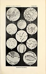
Plate VII Photomicrographs illustrating the more commonly encountered bacteria in European foulbrood. A. — Bacillus pluton: A smear from the stomach of a larva sick with European foulbrood. Note the paired forms and short chains. These forms are numerous in a recent infection, suggesting the organism in the process of multiplication. The lancet-shaped form is by far the predominant one in all later stages of the disease. X 1000. B. — Bacillus pluton: A smear from a larva quite recently infected. The multiplying paired forms are at this stage present almost exclusively. X 1000. C. — Bacterium eurydice: Stained preparation from a pure culture on the surface of agar. X 1000. D. — Bacillus alvei: Stained preparation showing spores and spore formation. X 800. E. — Streptococcus apis: Stained preparation from a pure culture. X 800. F. — Bacillus alvei: The peculiar arrangement of the spores as sometimes seen. From a pure culture, the smear having been made by suspending the culture on the slide in normal salt solution. X 1000. G. — Bacillus orpheus: Stained preparation made from a pure culture only a few hours old. Grown on the surface of agar. X 1000. H. — Bacillus orpheus: Stained preparation showing spore formation. Note the stained portion along one side and about both ends of the spore. The stage is soon reached in a culture at incubator temperature. At room temperature it remains in this stage for a considerable period. X 800. I. — Longisection of a young larva showing early infection in European foulbrood. The bacterial growth is seen as a narrow black area just within the peritrophic membrane on one side of the food mass. J. — Longisection of larva sick of European foulbrood. showing a later stage of infection than that present in I. The dark area in the food mass shows the bacterial growth. Note that the growth mass does not extend beyond the peritrophic membrane and that it does not extend uniformly along this membrane and throughout the food mass. K. — Transverse section of larva about the time of its death from European foulbrood infection. Note the bacterial mass along the peritrophic membrane and extending from the membrane into the food mass. As seen within the living larva this bacterial mass in the sick larva is practically white, but is more or less yellowish white when present with larval food material. The gelatinous-like envelope outside the peritrophic membrane and inside the stomach epithelium in healthy larvae thins out as the disease advances. |
||
| Fecha | |||
| Fuente | https://www.biodiversitylibrary.org/pageimage/41863898 | ||
| Autor | White, G. F. | ||
| Page ID | 41863898 | ||
| Item ID | 131054 (Find related Wikimedia Commons images) | ||
| Title ID | 64650 (Find related Wikimedia Commons images) | ||
| BHL Page URL | https://www.biodiversitylibrary.org/page/41863898 | ||
| DOI | 10.5962/bhl.title.64650 | ||
| Page type | Text Illustration | ||
| Credit | The contributing institution believes that this item is not in copyright
|

Licencia
Este archivo está en el dominio público porque es una exploración mecánica simple o fotocopia de un original en dominio público, o (con las pruebas disponibles) es tan similar a un documento escaneado o fotocopia que no se puede aplicar protección de derechos de autor. También puede suceder que los derechos de autor de esta imagen hayan expirado debido a la fecha de publicación o la muerte del autor (si es posible, añadirla aparte). El propio contenido original se encuentra en dominio público por las siguientes razones:
Esta etiqueta está diseñada para usarse cuando sea necesario afirmar que las mejoras (por ejemplo, brillo, contraste, juego de color, nitidez) no son de por sí suficientemente creativas para generar un nuevo derecho de autor. Se puede utilizar tanto cuando no se sabe si se han hecho mejoras como cuando las mejoras son claras, pero insuficientes. Para las imágenes primitivas sin contraste puede utilizar la plantilla {{PD-old}} adecuadamente insertada. Para utilizarla, véase Commons:Cuándo usar la etiqueta PD-scan.  | ||||
Historial del archivo
Haz clic sobre una fecha y hora para ver el archivo tal como apareció en ese momento.
| Fecha y hora | Miniatura | Dimensiones | Usuario | Comentario | |
|---|---|---|---|---|---|
| actual | 14:29 2 nov 2015 | | 2628 × 4260 (1,68 MB) | Fæ | == {{int:filedesc}} == {{BHL | source = http://www.biodiversitylibrary.org/pageimage/41863898 | description = European foulbrood / | pageid = 41863898 | itemid = 131054 | titleid = 64650 | pagenumbers = | pagetypes = Text Illustration | volume = no.8... |
Usos del archivo
La siguiente página usa este archivo:
Uso global del archivo
Las wikis siguientes utilizan este archivo:
- Uso en de.wikipedia.org